

Laminitis
Laminitis affects tissue called sensitive laminae which are found in the horse’s hoof. Laminitis occurs when blood supply to the hoof is compromised which may result in failure of the bond between the internal structures within the hoof. This can cause the pedal bone to move.
Thrush
Thrush is a common bacterial infection affecting the horse’s hoof, more specifically the soft frog tissues and sometimes the heel of the foot. It usually starts in the frog clefts and can produce black smelly discharge. It can affect one hoof or all four at a time.
White line disease
White line disease (also known as seedy toe) occurs when the hoof wall and sensitive laminae begin to separate at the white line. This causes a cavity which can then start to fill with dirt, horn and other debris exposing it to infection. Horn is the hard tissue which makes up the outer hoof wall. Once the horn has become compromised, anaerobic bacteria (meaning the bacteria don’t need oxygen to survive) begin to thrive and spread throughout the hoof wall.
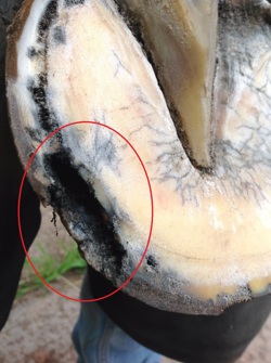
Photo credit: Nigel Brown AWCF
The highlighted area shows a large cavity caused by separation at the white line
Diagnosis and treatment
chevron-down
chevron-up
Diagnosis
When picking out the horse’s feet a noticeable cavity can be seen around the white line. The horn may also start to become crumbly. The condition doesn’t always cause lameness but if left untreated can become very serious.
If you’re concerned about your horse, contact your farrier or vet.
Treatment
A farrier may simply be able to cut back the affected area exposing the bacteria to oxygen, discouraging further growth. The area should be kept clean and dry to prevent further bacteria entering2. Your farrier or vet may apply iodine solution or other antiseptics to the area to aid in recovery.
In severe cases an x-ray may be needed to determine the degree of separation - a larger separation could cause the pedal bone to move within the hoof capsule resulting in laminitis.
Make sure that the horse regularly sees a farrier to maintain good hoof balance, this can help to prevent infection from occurring.
Penetrations
Penetrating hoof injuries are relatively uncommon in horses, but can be caused by nails and screws. Deep wounds can cause severe damage to the internal structures of the hoof causing permanent and severe lameness. Any penetrating injury should be immediately seen by a vet.
Bruised sole
This is one of the most common causes of lameness in horses3 and can affect both shod and unshod horses. Injury to the sole of the hoof can damage the sensitive structures within, causing bruising. Standing on a hard object such as a stone, repetitive work on hard ground or poor hoof care can all cause bruising.
Bruising occurs when the tiny blood vessels within the sensitive sole burst when damaged. Severe bruising can cause the formation of a ‘haematoma’3, this is like a blood blister and can be extremely uncomfortable for the horse.
Usually, the horse is only lame on the affected leg, which can be intermittent.
Diagnosis and treatment
chevron-down
chevron-up
The area of pain can be located by the vet or farrier using a pair of hoof testers. This area can then be carefully trimmed back which may reveal a red/purple bruise in white feet.
Bruises usually heal on their own, however, if severe bruising occurs the hoof may need to be poulticed with a protective bandage to relieve the weight-bearing pressure on that foot. Protective hoof pads may also be placed under the horse’s shoe to help prevent further bruising from occurring.
Nail bind
Nail bind describes the condition where a nail has been driven too close to the sensitive laminae during the shoeing process. Often this causes low degree pain and lameness, and there’s risk of an infection forming around the nail causing an abscess.
Diagnosis and treatment
chevron-down
chevron-up
Diagnosis
Signs of nail bind are often lameness following shoeing either immediately or a couple of days later. Heat and an increased digital pulse may also be present.
The problem nail can be determined using hoof testers.
Treatment
Usually, removing the impinging nail is enough to solve the issue. However, most of the time the whole shoe is removed, and a poultice applied to draw out any infection. A tetanus injection may be advised if the horse isn’t already vaccinated, as well as a course of antibiotics from your vet depending on the severity.
Abscesses
An abscess occurs when bacteria gets trapped between the laminae and hoof wall causing a localised infection. The infection builds a large amount of pressure within the hoof which causes sudden and severe lameness. Sometimes abscesses can burst out of the top of the coronet band if left untreated.
Changes in weather can cause the hoof to become dry and as a result can crack allowing bacteria into the hoof wall. Poor management can lead to abscesses if the hoof isn’t regularly trimmed, or if your horse suffers a penetration injury.
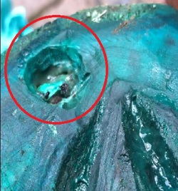
Photo credit: Nigel Brown AWCF
The image shows an abscess after an initial poultice being applied. The blue colour is an antiseptic which has been used by the farrier to draw out infection
Diagnosis and treatment
chevron-down
chevron-up
An abscess is often diagnosed using hoof testers to locate the specific area.
Abscesses can take varying lengths to heal depending on their severity. The vet or farrier will likely drain the area, relieving the pressure. It’s important to keep the area clean to prevent further infection spreading. Tubbing the hoof may be useful, this is where the horse stands in warm water with Epsom salts for approximately 15 minutes. Or a poultice can be applied to draw out any infections and keep the hoof clean.
Pain relief and antibiotics may be prescribed from your vet such as bute.
Grass cracks
Grass cracks tend to start at the bottom of the hoof or on the sole whereas sand cracks originate at the top of the hoof at the coronet band.
Grass cracks usually occur when the hoof becomes overgrown, and the pressure causes the horn to split or break off. This type of crack rarely causes severe lameness; however, it can lead to infections such as abscesses.
Sand cracks tend to occur due to poor foot balance or injury. They can be superficial, however if you notice a sand crack occurring it’s important to speak to your farrier as this often indicates that the horse may need trimming or shoeing differently.
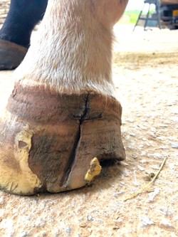
Photo credit: Nigel Brown AWCF
An untreated sand crack which has spread downwards
Treatment
chevron-down
chevron-up
Hoof cracks will not heal without intervention. It’s important to speak to your farrier to find the cause of the cracks before infection starts to take hold. It’s better to act early to prevent further damage to the hoof from occurring. Remedial shoeing or alternative procedures may be required to help correct and support the hoof while the crack grows out.
Find out more
Supported by Nigel Brown AWCF on behalf of the British Farriers and Blacksmiths Association.
![]()
References
- Belknap, J. (2022) Disorders of the Foot in Horses.
- Main Street Veterinary Clinic. (2016) Seedy Toe.
- Carson, D. & Ricketts, S. (2022) Bruised Sole in Horses.
Donkeys
Donkeys are also susceptible to similar hoof issues. For specific advice visit The Donkey Sanctuary.