External structures (points of the hoof)
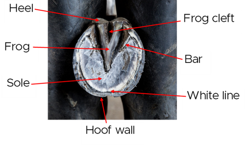
Hoof wall
chevron-down
chevron-up
The hoof wall is the hard outer layer that protects everything inside the hoof. It’s made from keratin, the same material as human hair and nails. The wall grows slowly from the top (coronary band) to the toe, taking about 9–12 months1. The wall supports most of your horse’s weight and plays a critical role in maintaining balance and stability. A healthy hoof wall should look smooth, without any cracks, lumps or lines. The hoof can be one solid colour (often brown or white) or have stripes of different colours.
Coronary band
chevron-down
chevron-up
The coronary band is the strip at the top of your horse’s hoof where the skin meets the hoof wall. This area creates new hoof growth and sends nutrients to the wall. If the coronary band is damaged, it can affect how the hoof grows.
Sole
chevron-down
chevron-up
The sole is the bottom part of the hoof. It’s slightly curved (concave) to help absorb shock and protect the inside of the hoof. It’s made of keratinized tissue that provides a firm yet flexible barrier against ground pressure. While strong, it’s not designed to bear full weight, which is primarily supported by the hoof wall.
Frog
chevron-down
chevron-up
The frog is the soft, V-shaped part in the middle of the hoof’s underside. It helps absorb shock and pushes blood back up the leg when your horse moves. It’s important for healthy circulation and hoof flexibility.
Bulbs of the heel
chevron-down
chevron-up
The heel bulbs are the rounded, soft parts at the back of the hoof. They allow the hoof to expand and contract when your horse moves. This helps absorb impact and keeps the hoof stable on uneven ground.
White line
chevron-down
chevron-up
The white line is where the hoof wall and sole meet. It holds the hoof together and acts as a barrier to keep out dirt and germs. Problems can happen if this line becomes weak or stretched.
Internal parts
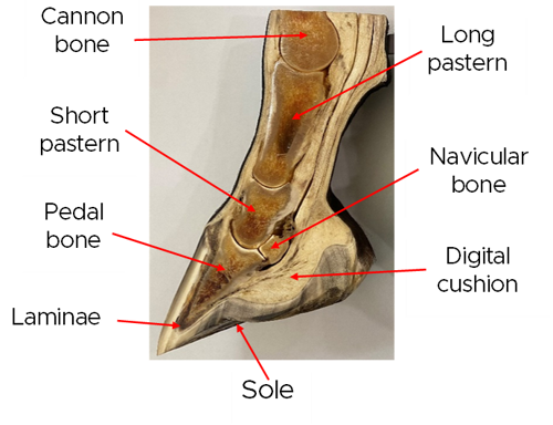
Pedal bone (coffin)
chevron-down
chevron-up
The pedal bone is the main bone inside the hoof. It has a strong, slightly curved shape that fits the hoof and helps spread your horse’s weight evenly. This bone supports their movement and connects to important structures like tendons and ligaments. Its position and health are vital for balance and soundness.
Laminae
chevron-down
chevron-up
The laminae are thin, leaf-like layers inside the hoof. They attach the hoof wall to the pedal bone, working like strong hooks to hold everything in place. There are two types: sensitive laminae (with blood supply) and insensitive laminae (part of the hoof wall). Together, they keep the pedal bone secure and allow the hoof to grow and function properly.
Digital cushion
chevron-down
chevron-up
The digital cushion is a thick, soft pad located under the pedal bone and behind the frog. It acts like a shock absorber when the horse moves, reducing impact on bones and joints. It also helps pump blood back up the leg, which is important for circulation and hoof health2.
Collateral cartilages
chevron-down
chevron-up
These are two pieces of cartilage on each side of the hoof, near the heel. They help the hoof expand and contract with movement, making it more flexible. They also protect the joints inside the hoof and support smooth, natural motion.
Navicular bone (distal sesamoid)
chevron-down
chevron-up
The navicular bone is small and sits behind the pedal bone, inside the hoof. It’s held in place by strong ligaments and cushioned by a fluid-filled sac called the navicular bursa. This sac allows the deep digital flexor tendon to slide smoothly over the bone3. Both the bone and the bursa are part of the joint between the pedal bone and short pastern (often called the coffin joint), which plays a key role in movement and weight-bearing.
References
- Pollitt, C. (2015) The Illustrated Horse's Foot.
- Kauffman, S. & Cline, C. (2018) The Essential Hoof Book.
- Al-Agele, R. et al., (2019) The Anatomy, Histology and Physiology of the Healthy and Lame Equine Hoof.